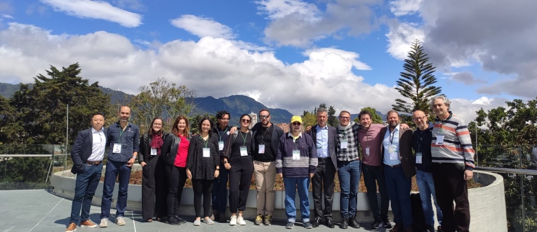
NEU-ChiP visits Sicily

Presenting the consorta at the 2023 International Symposium on Nonlinear Theory and Its Applications (NOLTA) a number of the group were able to present over the week event, bring in lots of discussion and interest from others, as well as push forward ideas amongst NeU-ChiP partners in various places, even on Mount Etna. Papers and abstracts from the event can be found here.

A special symposia for NEU-ChiP ran over two days as a hybrid meeting, enabling discussion with collaborators around the world.

Around the physical sessions the NEU-ChiP team found time to discuss ideas.

Around the conference we had the chance to see a little of the city of Catania, with some great food, and an excellent conference dinner in the beautiful museum.

The NEU-ChiP team also found some time to enjoy the surrounding sites with some adventures up to Mount Etna.

Blog – Mathematical Modelling the Brain

The brain is one of the most complex organs in the human body, responsible for everything from our thoughts and emotions to our ability to move and sense the world around us. It is a fascinating and mysterious structure, and scientists have been studying it for centuries in an attempt to understand how it functions.

One of the most recent and exciting developments in this area is the use of mathematical models to understand the brain. Mathematical models are simplified representations of complex systems, and they can be used to predict the behavior of those systems under different conditions.

In the context of the brain, mathematical models can help us understand how neurons communicate with each other, how neural networks form, and how the brain processes information. They can also be used to simulate the effects of drugs or other interventions on the brain, which could lead to the development of new treatments for neurological disorders.

One of the most famous examples of a mathematical model of the brain is the Hodgkin-Huxley model, developed in the 1950s. This model describes the behavior of neurons and their ability to transmit electrical signals. Since then, many other mathematical models have been developed, each one building on the knowledge gained from previous models.

One of the key advantages of using mathematical models to study the brain is that they allow us to explore the behavior of the brain in a way that would be impossible with traditional experiments. For example, it would be difficult to study the behavior of millions of neurons in real-time, but a mathematical model can simulate this behavior and allow us to explore the consequences of different scenarios.

Mathematical models can also be used to test hypotheses in a more systematic way. Instead of relying on trial-and-error experiments, researchers can use mathematical models to predict the outcome of an experiment before it is conducted. This can save time and resources and lead to more efficient research.

Of course, there are also limitations to using mathematical models to study the brain. For example, mathematical models are only as good as the data that goes into them, and there is still much we don’t know about how the brain functions. Additionally, mathematical models can only provide a simplified representation of the brain, and it is important to remember that they are just one tool in the arsenal of neuroscientists.

In conclusion, the development of mathematical models to understand the brain is an exciting and rapidly evolving field of research. By using these models, scientists are gaining new insights into how the brain functions and how it can be treated when it malfunctions. While there are limitations to using mathematical models, their potential for advancing our understanding of the brain is enormous, and we can expect to see many more exciting developments in the years to come.

Blog – Living cell circuits

Creating Living Circuits with Microfabrication

Microfabrication tools have revolutionized the way we engineer biological systems, allowing us to manipulate living cells at a level of precision that was once impossible. Among the many applications of microfabrication in the field of biology is the creation of living circuits from neurons in vitro. This technique has the potential to revolutionize the field of neuroscience, by providing a platform for studying the behavior of neurons and neural networks in a controlled environment.

In this blog post, we will explore how microfabrication tools can be used to create living circuits from neurons in vitro, and discuss some of the potential applications of this technique.

What are living circuits from neurons in vitro?

Living circuits from neurons in vitro are networks of neurons that are grown in a dish and connected in a specific pattern using microfabrication tools. These circuits can be used to study the behavior of neurons in a controlled environment and to explore the properties of neural networks.

The basic idea behind living circuits is to create a pattern of microchannels on a substrate, which can be filled with a solution containing neurons. The neurons then grow in the microchannels, forming connections with each other and creating a functional network.

The process of making living circuits from neurons in vitro involves several steps, including microfabrication, cell culture, and network formation. Let’s look at each of these steps in more detail.

Microfabrication: The first step in making living circuits is to create a pattern of microchannels on a substrate. This is typically done using photolithography, a technique that uses light-sensitive materials to create patterns on a substrate. The substrate can be made of a variety of materials, including glass, silicon, or polymer.

Cell culture: Once the microchannels are created, the next step is to culture neurons in the channels. This is done by seeding the channels with a solution containing neurons. The neurons will adhere to the surface of the channels and begin to grow.

The final step is to allow the neurons to form connections with each other, creating a functional network. This is typically done by allowing the neurons to grow for several days or weeks, during which time they will form connections with each other and begin to communicate.

Living circuits from neurons in vitro have many potential applications in the field of neuroscience. One potential application is in the study of neurodegenerative diseases, such as Alzheimer’s and Parkinson’s disease. By creating living circuits from neurons in vitro, researchers can study the effects of these diseases on the behavior of neurons and neural networks, which could lead to the development of new treatments.

Another potential application of living circuits is in the development of neural prosthetics. Neural prosthetics are devices that can be implanted in the brain to restore lost function, such as the ability to move or communicate. By studying the behavior of neurons in living circuits, researchers can develop better prosthetics that are more effective and longer-lasting.

In conclusion, living circuits from neurons in vitro are an exciting new tool for studying the behavior of neurons and neural networks. By using microfabrication tools to create these circuits, researchers can study the effects of disease, develop new treatments, and create better neural prosthetics. With continued research and development, the potential applications of living circuits are endless, and we are only just beginning to scratch the surface of what is possible.

Blog – Undergraduate MChem Student at Loughborough University, UK

I’m an Undergraduate Master of Chemistry student at the University of Loughborough in my final year, due to graduate in the summer of 2023. During my time at university, I have performed investigations into the impact of differing concentrations and combinations of the chemotherapy drugs Paclitaxel and Fluorouracil on the neuroblastoma SH-SY5Y cell line, the effectiveness of Ampicillin and Chloramphenicol on the bacteria E. coli and S. epidermidis, as well as many other organic synthesis and analytical investigations. I have previously written research literary reviews on the utilisation of microfluidics-based blood-brain-barrier models to understand and treat glioblastomas, and the recreation of oriented neural networks on a chip through probabilistic guidance of axonal growth – the latter being the area I am focusing my novel NeuChip investigation on.

After doing my first Biochemical experiments last year, I realised that it was the area of chemistry I was most interested in and wanted to pursue the medicinal and pharmaceutical aspects of biochemistry with the study of in vitro models also.

“Dan is a great student and a pleasure to have in the team. I’m looking forward to following his career trajectory as he is so motivated and enthusiastic – he will certainly go places and do amazing things.”

Dr Paul Roach, Loughborough University

You can find out more about Dan in his profile video here.

Brains in a Dish Artwork Display

Dr Eric Hill and colleagues from Aston enjoyed an “exciting new immersive exhibition” to support the open delivery and accessibility of science to the community through large scale kinetic installations with glass, light and body recognition software.

This is an accumulation of over 7 years work, with an amazing online gallery here.

Visual Display of Neurons

David Jenkins yet again shows off the multi-couloured delights of his neurons in a linked video here. Two of his fellow researchers are partially colour blind, so to help with their visualisation of the cell types and domains he has selected colours that they can discriminate.

See more of the amazing images in the NEU-CHiP gallery.

Summer Placement in Lboro NEU-ChiP Labs

Loughborough School of Science hosted 3 excellent students and 2 amazing teachers from St. John’s College Cardiff over a week in the summer. Everyone had hands on experience of multiple lab practicals ranging from culturing living cells to making micro-devices in a clean room. The students fed back from their experience that they learned a lot and were able to explore areas of science that they have never thought about.

The students got to grips with the micro world, using a scanning electron microscope to see the surfaces of materials at this very small scale.

During July, I undertook a work experience with Dr Roach at Loughborough University which was an incredible experience. It really brought my passion for science alive and I was able to experience chemistry and biology in a practical setting. My most memorable moment was probably gas chromatography which interested me by how many uses it had and the explanations by the members of staff were brilliant. I was able to use this experience to strengthen my application for medicine and it motivated me to continue along this career path.

Matthew Logan

Using Schlieren photography you can visualise small changes in air density, even the thermal changes from a hand.

We went to Loughborough University to undertake work in the laboratories there with Dr. Roach for work experience, during which we experienced quite a lot of amazing things that combined chemistry, physics and biology together. My favourite part was looking at electron microscopy, as I could look into any part I wanted to observe and the appearance was so magical that it let me realize that a very, very small corner on the earth could have such a wonderful world inside. Besides this we also done many fantastic things such as cell cultures, fluorescent imaging and analysis Chemistry which greatly broadened my horizons. As an overseas student, I haven’t done any formal practical work before the AS level, so this trip was quite challenging for me, but it really taught me a lot of useful skills and developed my interest in natural science. Really want to thank Dr. Roach as he was so kind and knowledgeable that he arranged our rewarding trip perfectly and helped us so much along the way.
Thank you very much for this amazing trip!

Estella Shi

It was quite a long week with lots to see and do, but everyone left smiling and making plans for a return next year. St. John’s College Cardiff students are welcome anytime – and their teachers are pretty fun too.

Paul Roach